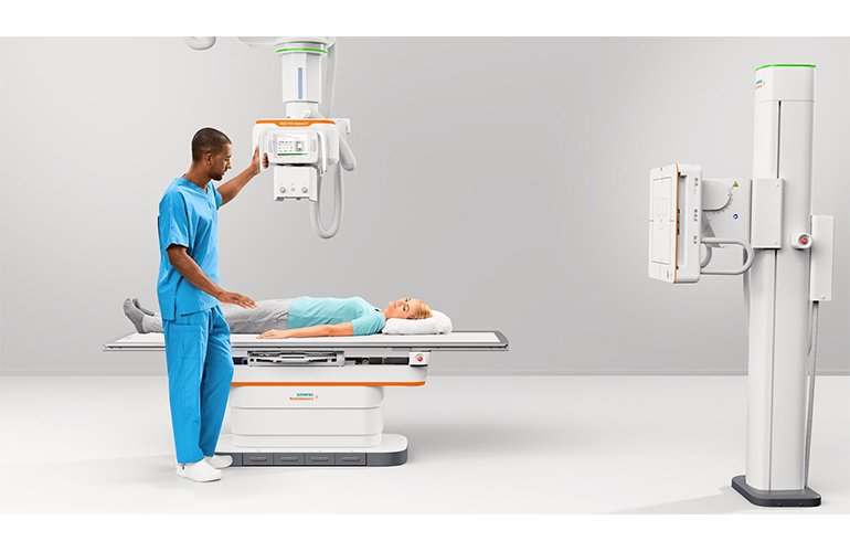
- Digital X-rays
- C.T. Scan
- Sonography and Color Doppler (Peripheral and Carotid)
- Examination of female patients by a female sonologist


DIGITAL X-RAY
Digital X-ray sensors are used instead of traditional photographic film. Its advantages include time efficiency through bypassing chemical processing and the ability to digitally transfer and enhance images.
Radiography with X-ray is the key starting point for diagnosing a variety of health issues, including pneumonia. It is a technique listed under the domain of Digital radiography. X-ray technology is easily available to the medical community because of its low cost. It’s absolutely noninvasive, harmless if not overdone, and produces the images quickly so that we can be able to diagnose issues effectively.
C.T. Scan
A CT scan uses computers and rotating X-ray machines to create cross-sectional images of the body. These images provide more detailed information than typical X-ray images. They can show the soft tissues, blood vessels, and bones in various parts of the body.
A CT scan may be used to visualize the:
- head
- shoulders
- spine
- heart
- abdomen
- knee
- chest


Sonography and Color Doppler (Peripheral and Carotid)
Healthcare providers use Doppler ultrasound to detect heart and blood vessel (cardiovascular) problems. The test shows the direction and speed of blood moving through arteries and veins. It can identify blood clots, narrowed arteries, and other problems that affect the heart and blood vessels in the legs, arms, and stomach.


